
1 week ago
Marsico Lung Institute/UNC Cystic Fibrosis Center
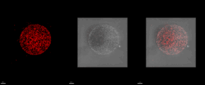
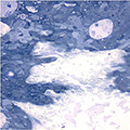
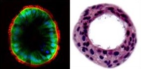
Pseudomonas aeruginosa, a source of lung infection in cystic fibrosis.
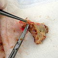
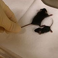
The images show Pseudomonas aeruginosa bacteria, MPAO1, expressing a red fluorescent protein embedded in an agar bead visualized by confocal microscopy. Fluorescent P. aeruginosa (left); phase contrast (middle); merged image (right). These beads are delivered into the airways of mice and are used to develop a chronic infection for up to 28 days. With this novel chronic infection model, we are able to better investigate bacterial and host determinants driving morbidity and mortality in cystic fibrosis.
These images are from the Wolfgang Lab and were taken by Matthew Greenwald, PhD.
Recent News

2 months ago
Marsico Lung Institute Publications for November 2025

2 months ago
Marsico Lung Institute Publications for October 2025
3 months ago
Sun Featured as Emerging Investigator in October AJRCCM